Bitemarks (Orthognathic 3D Splints)
Specialist in Cranio-Maxilofacial Surgery & Customized Implants
Bitemarks
Modern 3D virtual planning for orthognathic surgery has critical advantages compared to conventional treatment planning. In the traditional method, in order to achieve a precise diagnosis of the dentoskeletal deformity and create a treatment plan intraoperatively reproducible, it is necessary to collect data from different sources, such as photographs, cephalograms, dental casts, physical examination along with face bow record and its transfer to semi-adjustable articulators, and measurement of plaster casts movement, according to the surgical simulation.
Simulating the operation on plaster dental casts is difficult, especially in cases of complicated two-jaw surgery, as it requires many laboratory-based steps that are time-consuming and may lead to potential errors.
As a result of developments in 3D imaging technology, surgeons are provided with extra information that could not be obtained from lateral cephalogram alone, thus improving the quality of preoperative planning.
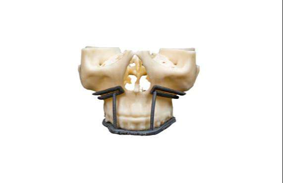
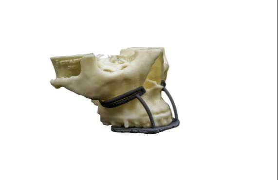
3D printing technology allows us to replicate more closely the actual patient while providing access to more and higher-quality information about patient’s 3D anatomy and improving the ability to identify conditions that are not detectable with 2D conventional imaging techniques, thus improving the accuracy and reliability of diagnosis and treatment.
Moreover, unlike conventional model surgery on dental casts, this technology allows to virtually perform multiple simulations of different osteotomies and skeletal movements in order to evaluate multiple surgical plans.
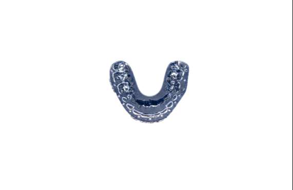
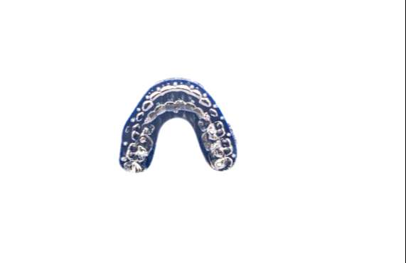
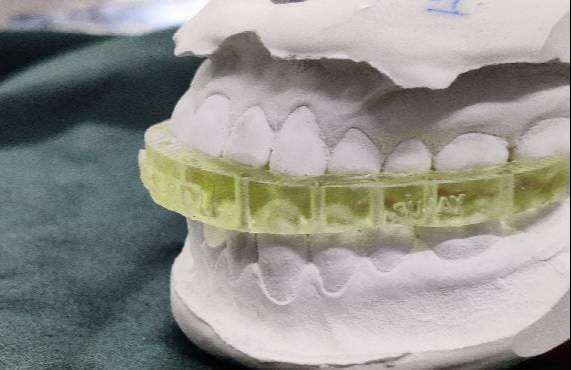
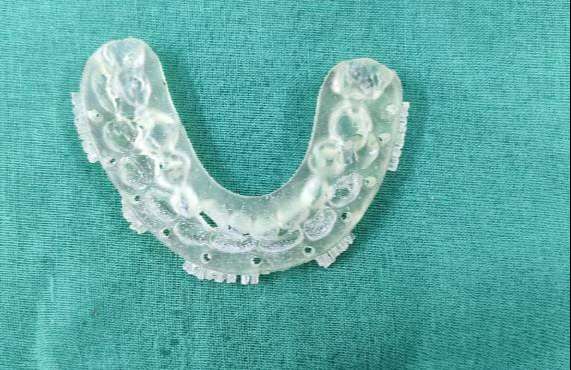
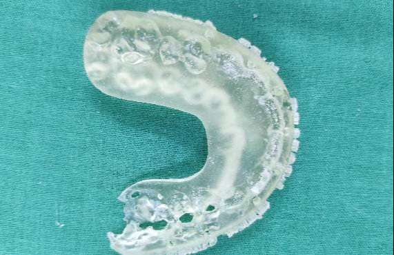
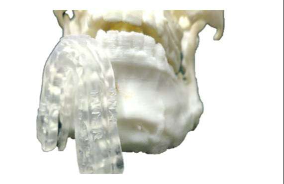
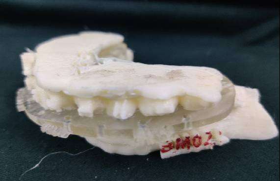
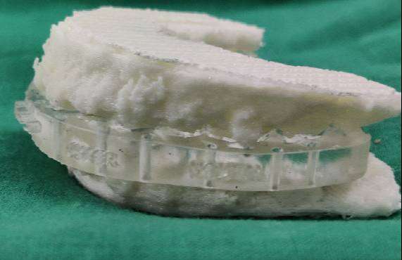
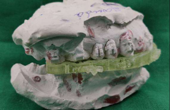
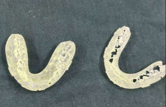
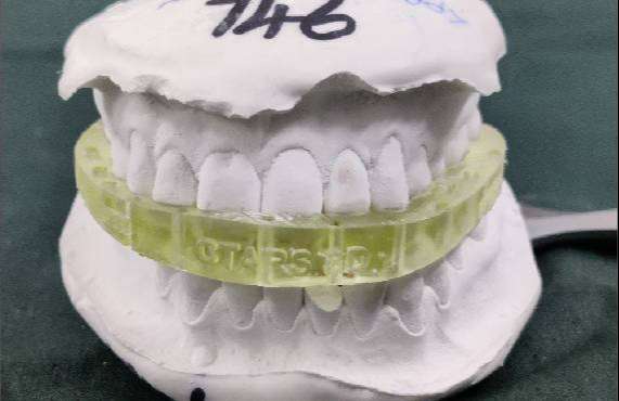
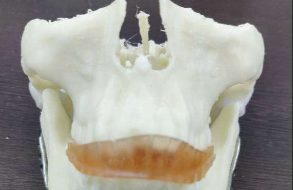
At CTARS, the Orthognathic splints that we produce are made up of polyjet and printed using 3D Polyjet technology.


